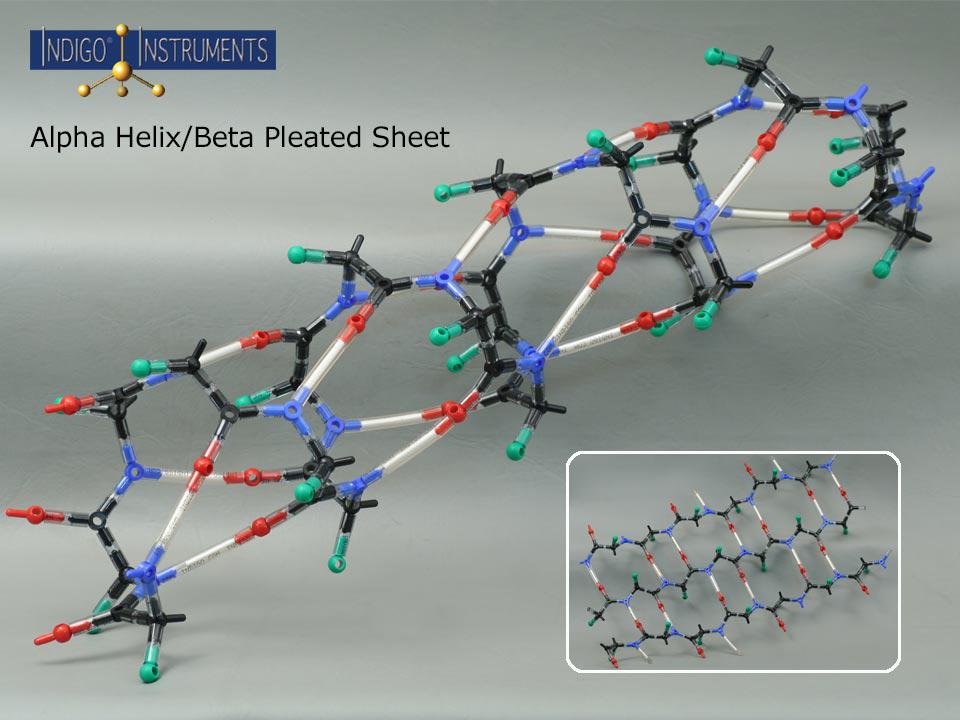
This Indigo® molecular model kit can build an alpha helix or beta sheet to explore hydrogen bonding, sidechain orientation and more. Use it for understanding Protein Secondary Structure and a Biochemistry Protein Folding Teaching Tool.
This molecular model kit provides a tangible way to visualize hydrogen bonding, backbone geometry, hydrogen bonding and sidechain orientation in an alpha helix. The open framework of this model highlights the intralocking, intra-chain hydrogen bonds that hold the secondary structure together. It also fits the major groove of our 12-base-pair Indigo® DNA double helix model making it an ideal companion teaching tool. By manipulating the model, learners can explore how amino acid sequence drives structure and gain a hands-on understanding of protein folding, stability, and interactions
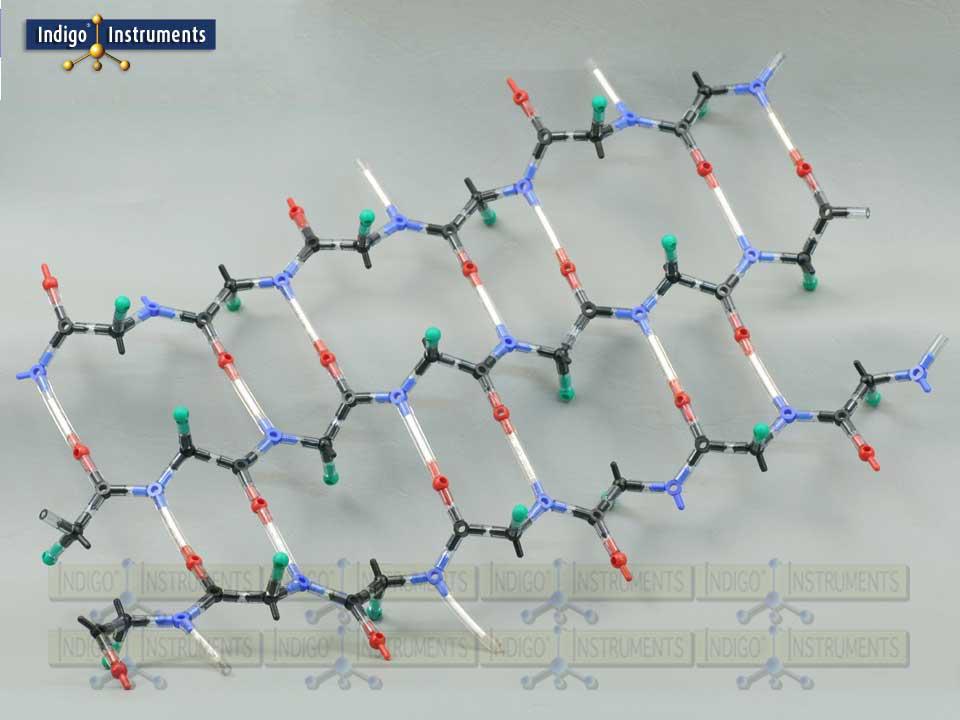
“R” groups are indicated with a green "atoms" but parts that represent actual sidechains can be added. The 50mm bonds with white inserts represent the amide hydrogen atoms (N–H) of residues that form hydrogen bonds with the carbonyl oxygen atoms (C=O) of another residue. In alpha helices, these bonds stabilize the coiled structure with the characteristic i → i+4 pattern, while in beta sheets, hydrogen bonds form between strands in either parallel or antiparallel arrangements. This makes it easier to visualize how hydrogen bonding contributes to the stability and geometry of both secondary structure types.
The hydrophobic side chains of the constituent amino acids of an alpha helix interact with the hydrophobic interior of the lipid bilayer which stabilizes its position within the cell membrane.
As transport proteins, alpha helices act as pathways to allow specific ions to move across cell membranes. These channels can be selective for water or act as gates for specific ions such as sodium, potassium, and calcium.
Bundles of alpha helices that span cell membranes change their conformation in response to ligand binding which in turn initiates intracellular signalling pathways. Some may even detect changes in light or pH & play a role in photosynthesis and respiration & electron transport as part of energy transduction.
Circular dichroism (CD) spectroscopic analysis can inform the secondary structure, stability, and conformational changes in alpha proteins through comparing the interaction of polarized light the arrangement of peptide bonds. Alpha helix protein spectroscopic features peak in the far UV region of 190-250nm. Changes in the UV spectrum can help quantify a number of conditions affecting protein structure. This includes the extent of alpha helical content in a protein, the degree of its folding vs unfolding in response to temperature & pH or the binding of ligands.
Indigo Instruments has held inventory of genuine Cochranes of Oxford (Orbit) parts for 30+ years (See Skeletal (Orbit/Minit)) that are compatible with every molecular model we have sold since day 1. This level of quality may appear expensive but no parts support from other vendors costs even more.
| Concept | Observable / Hands-On Outcome | Applicable Audience |
|---|---|---|
| Alpha helix hydrogen bonding | Identify i → i+4 H-bonds in a coiled helix | Biologist, Biochemist |
| Beta sheet strand pairing | Build parallel vs antiparallel sheets and observe H-bond patterns | Biologist, Biochemist |
| Sidechain orientation | Visualize how R-groups project above and below the plane of the sheet or helix | Biochemist, Drug Designer |
| Sequence → structure relationship | See how amino acid patterns stabilize helices or sheets | Biologist, Biochemist, Drug Designer |
| DNA interaction | Explore how the alpha helix fits the major groove of DNA | Biologist, Biochemist |
| P/N | Description | QTY |
|---|---|---|
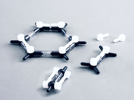
| 67288c | Kit Use Only Bond, Minit, white, 210mm | 1 |
| 68186-20 | Wobbly bond, 20mm, each | 100 |
| 68186-50 | Wobbly bond, 50mm, each | 17 |
| 68221C | Atom, Orbit, Cl "a", green, 1 prong | 20 |
| 68227C | Atom, Orbit, O "b", 180 degree, red | 20 |
| 68236C | Atom, Orbit, C "h", planar: 108-126-126, black | 20 |
| 68237C | Atom, Orbit, N "h", planar: 108-126-126, blue | 20 |
| 68244C | Atom, Orbit, C "k", tetrahedral, black | 20 |
Sidechains can be added to show how amino acid variation influences the stability and properties of secondary protein structures. The sidechains shown in the table are for illustrative purposes and are equally applicable to the alpha helix and beta sheet.
| Side Chain | Methionine | Alanine | Glutamate | Lysine |
|---|---|---|---|---|
| Part | ||||
| 68216c | 7 | 3 | 5 | 10 |
| 68218c | 1 | |||
| 68228c | 1 | |||
| 68230c | 1 | |||
| 68241c | 1 | |||
| 68244c | 3 | 1 | 2 | 4 |
| 68245c | 1 | |||
| 68186-20 | 7 | 3 | 5 | 10 |
| 68186-30 | 4 | 1 | 4 | 5 |
| 68267c | 2 | |||
| 68366 | 1* |
*The glutamate side chain uses 1 of 15 bonds in the bundle to connect 2 X-pegs for the C=O double bond.
For each Related Product, enter the quantity you would like to order and click the Add to Cart button beside the item.
Methionine (met), alanine (ala), leucine (leu), glutamate (glu), and lysine (lys) are most common in alpha helices. Proline (pro) because its ring's structure has steric hindrance and glycine's small, flexible side chain make them both very rare.
An α-helix is a coil of amino-acid residues in a polypeptide chain. This helical coil is held together by hydrogen bonds between a carbonyl oxygen (C=O) and an amide hydrogen (N-H) 3 amino acid residues below it in the chain sequence.